Hospitals eTool
Surgical Suite » Ionizing Radiation Exposure
Hazards
Staff are exposed to ionizing radiation from radiation-generating devices used during surgical procedures. Examples include X-ray machines and fluoroscopy units.
Exposure
Radiation dose is received when workers are near an operating radiation-generating device or a radioactive source. The dose received depends on the type of radiation, the amount of radiation generated, the duration of exposure, the distance from the source of radiation, and the amount and type of shielding in place.
Health Effects
Surgical staff members may be repeatedly exposed to low levels of ionizing radiation over the course of their careers. Adverse health effects, such as cancer, may occur years following such exposure. The probability of an adverse health effect occurring is proportional to the radiation dose received (World Health Organization 2016).
Studies of atomic bomb survivors have shown significant associations between cancer and radiation dose levels of about 10 rems (0.1 Sv) or greater, with the cancer risk increasing as the radiation dose increases. For low-level radiation exposure (i.e., whole body doses less than about 10 rems (0.1 Sv)), statistical limitations in studies have made cancer risk assessment more difficult (National Research Council et al. 2006).
In 2006, the National Research Council's Committee to Assess Health Risks from Exposure to Low Levels of Ionizing Radiation reviewed the available data and concluded that the cancer risk would continue linearly at low doses. This finding means that there is likely no safe exposure level (i.e., threshold) and that even low radiation doses have the potential to cause a small increase in cancer risk (National Research Council 2006).
In addition to cancer, cataracts (i.e., detectable lens opacities) are another radiation-induced health effect that could occur in surgical staff (International Commission on Radiological Protection 2011).
For more detailed information on health effects from radiation exposure, see OSHA's Ionizing Radiation Safety and Health Topics Page.
For OSHA requirements on Ionizing Radiation (e.g., exposure limits and monitoring, posting, and recordkeeping requirements), see OSHA's Ionizing Radiation Standard (29 CFR 1910.1096).
Dose Monitoring
For OSHA requirements on personnel monitoring, see OSHA's Ionizing Radiation Standard (29 CFR 1910.1096).
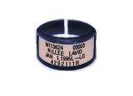
Personal dosimeters can be used for the long-term monitoring of a worker's radiation dose. Dosimeters include thermoluminescent finger dosimeters (TLDs), optically stimulated luminescence body (OSL) dosimeters, or film badges.


These passive dosimeters for personal exposure monitoring can be worn whenever working with radiation-generating devices, radioactive materials, or radioactive patients. Depending on the work situation, wear a body dosimeter at collar level, chest level or waist level. Wear finger ring dosimeters on the hand which is closest to the radiation source.
Personnel who work in high-dose fluoroscopy settings may be asked to wear two dosimeters for additional monitoring. Oftentimes, one dosimeter is worn on the outside of the lead apron at the collar (unshielded) and one on the inside at the waist (shielded).
Other Radiation Safety Practices
Provide radiation safety training to all workers who operate or are exposed to radiation-generating equipment, radiation sources, or radioactive materials.
Keep radiation exposures As Low As Reasonably Achievable (ALARA), and certainly below regulatory limits.
The three basic concepts of radiation protection are: (1) minimize the time of exposure, (2) maximize the distance from the source of radiation, and (3) use shielding. Applying these concepts will help to keep radiation exposures ALARA.
Some examples of radiation protective practices include:
- Using the shortest practical irradiation times.
- Using radiation-absorbing shields such as ceiling-suspended lead shields and table-suspended drapes as barriers to protect against X-ray exposure when procedures are in close proximity to the patient.
- Providing and ensuring employees use personal protective equipment (PPE) lined with lead or lead-equivalent materials (e.g., products made from dense elements or alloys of Pb, Sb, Bi), including lead or lead-equivalent aprons, thyroid collars, leaded eyewear with protective side shields, and leaded gloves for use by workers in the X-ray field. Ensure that workers do not place their hands in the primary X-ray beam. Providing and ensuring employees use properly fitted aprons to reduce ergonomic hazards and provide optimal radiation protection (Miller et al, 2010).
- Ensuring that procedures, like those that use remote fluoroscopy, are run using controls in an adjacent room, to the extent feasible.
- Giving a specific person the responsibility for ensuring proper maintenance of portable X-ray machines. Preventive and corrective maintenance programs for X-ray machines are detailed in 21 CFR Part 1000, Radiological Health, U.S. Food and Drug Administration.
- For detailed guidance on recognized practices, see:
- Federal Guidance Report No. 14. Radiation Protection Guidance for Diagnostic and Interventional X-Ray Procedures (FGR14). U.S. Environmental Protection Agency Interagency Working Group on Medical Radiation. November, 2014.
- Miller DL, et al. Occupational Radiation Protection in Interventional Radiology: A Joint Guideline of the Cardiovascular and Interventional Radiology Society of Europe and the Society of Interventional Radiology. Cardiovasc Intervent Radiol, (2010). 33:230-239.
- National Council on Radiation Protection and Measurements (NCRP), Report No. 168, Radiation Dose Management for Fluoroscopically-Guided Interventional Medical Procedures, (July 21, 2010).
- National Council on Radiation Protection and Measurements (NCRP), Report No. 105, Radiation Protection for Medical and Allied Health Personnel, (September 15, 1989).
- National Council on Radiation Protection and Measurements (NCRP), Report No. 102, Medical X-ray, Electron Beam, and Gamma-ray Protection for Energies up to 50 MeV, (April 4, 1989).
- Note that additional controls are required for the use of radioactive materials in hospitals, as regulated by the Nuclear Regulatory Commission. See 10 CFR 20, Standards for Protection against Radiation and 10 CFR 35, Medical Use of Byproduct Material.
Additional Information
- Health Risks from Exposure to Low Levels of Ionizing Radiation: BEIR VII, Phase 2. National Research Council, (2006).
- Ionizing Radiation. OSHA Safety and Health Topics Page.
- Ionizing Radiation Fact Book. U.S. Environmental Protection Agency (EPA). The Offices of Air and Radiation and of Radiation and Indoor Air published this document, which includes information on the health effects of ionizing radiation.
- Ionizing radiation, health effects and protective measures. World Health Organization (WHO), (April 2016).
- OSHA Technical Manual (OTM) - Hospital Investigation Health Hazards, Section VI: Chapter 1.
- Statement on Tissue Reactions. International Commission on Radiological Protection (ICRP), (April 21, 2011).
- Webster, E.W. EDE for Exposure with protective aprons. Health Physics Journal 1989. 56(4):568-569.
- Memorandum of Understanding between the U.S. Nuclear Regulatory Commission and the Occupational Safety and Health Administration. OSHA, (September 6, 2013). Clarifies areas in which each Agency has jurisdiction.

